
Theoretical physicist Dan Rokhsar leads a double life.
Some days, he applies himself to traditional physics -- for now, the theory behind strange phenomena in supercold clouds of atoms called Bose-Einstein condensates. When he can, though, he thinks about the brain.
Rokhsar is one of a growing number of Berkeley chemists and physicists applying the tools of their field to problems in biology.
And biologists are thrilled.
 Neuroscientists Corey Goodman and Carla Shatz want to rope physical scientists like Rokhsar and a broad range of researchers -- geneticists, psychologists, vision scientists and computer scientists -- into a center that would leapfrog our understanding of the brain. A new neuroscience center is one of Berkeley Chancellor Robert Berdahl's priorities, and a priority of the campus's New Century Campaign, which aims to raise $1.1 billion by the year 2001.
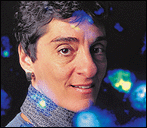
Rokhsar is trying to explain the ghostly images that flit across the retina during the eye's early development. Shatz has shown that these images -- generated spontaneously in the fetus -- are critical to establishing the proper nerve connections between the eye and the brain. Without them, the eyes are unable to see after birth.
"Dan and his student, Dan Butts, have developed beautiful models that turn out to have broad significance," Shatz said. "It's great that he's working to bring physics into biology."
If Goodman and Shatz have their way, collaborations like these would become routine.
"Corey and I both have a real vision that the Neuroscience Center we would build on campus would be a place not just where the best current neuroscience research would be going on, but also where people would be pushing the technical envelope that is really stopping the progress of our understanding of human brain function," Shatz said.
Though former President George Bush proclaimed this the decade of the brain in 1990, the 20th century ends with a big hole in our understanding: how individual nerve cells are assembled into such an amazing organ. Neuroscientists have elucidated many aspects of memory and vision, and have shown how the cells connect between the senses and the brain, as well as within the brain. But a top-to-bottom understanding remains elusive.
Imaging technologies like MRI (magnetic resonance imaging), CT (computed tomography) and PET (positron emission tomography), for example, have allowed scientists to "light up" regions of the brain that are active when we make decisions, see a pattern or engage in other activities.
Yet these regions represent millions of cells. Neuroscientists like Shatz study groups of 10 or 100 neurons.
"One wants to use imaging techniques to build a bridge between what we know about the functioning of a few neurons in a simple brain circuit to the functioning of the whole ensemble in a human. It is a huge gap in our knowledge right now," she said.
Goodman and Shatz, investigators in the Howard Hughes Medical Institute, are eager to draw in physicists and chemists to help in developing new techniques.
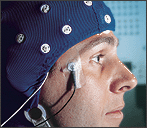
One such technique involves sensitive magnetic field detectors called SQUIDs (superconducting quantum interference devices), which have been incorporated into supercooled helmets that can measure the faint electrical signals sparking through the brain. Berkeley physicist John Clarke, a world expert on SQUIDs, says they can provide millisecond movies of brain activity, something no other brain scanning device can do.
"They've improved to the point where exciting things will begin to happen in studies of the brain," he said.
On the other hand, MRI shows exquisite detail. The biggest drawback is that MRIs require massive magnets. Alex Pines, a campus chemist who has refined MRI to an art, is working on improvements that could rid these machines of unwieldy magnets in favor of small or even no magnets.
Imaging machines like these are the bread and butter of Richard Ivry, a Berkeley professor of psychology who has joined with Goodman and Shatz to push for closer ties between cognitive scientists and neuroscientists. These machines are essential in pinpointing the areas of the brain that juggle specific tasks. Ivry's main interest is coordinated movement, critical in functions as simple as walking.
"We ask questions about how the brain controls the body parts -- the legs, the arms, the fingers -- but also, at a more abstract level, how we choose to make one particular action versus another," Ivry said. Such studies not only provide a better understanding of how the brain works in healthy people and patients with neurological disorders, but also can provide better strategies for coping with disease.
Ivry works closely with neurologists at the Veterans Administration Medical Center in Martinez to identify patients with damage in specific areas of the brain because of stroke, tumors or surgery.
"Our collaboration with the VA neurologists has allowed us to become a great department for patient-oriented studies," he said.
One current study involves split brain patients: people, often epileptics, who have undergone surgery to cut the corpus callosum, the main relay between the left and right hemispheres. Recent studies of such patients hint that the cerebellum -- a portion of the midbrain important in coordination -- acts as an internal timing system. Because split-brain patients have essentially two separate cortexes, they can control their hands independently -- they can simultaneously draw different figures with each hand, for example. Yet they do so with a synchrony that suggests there is an internal rhythm, perhaps provided by the cerebellum, which remains intact.
"Though I don't have conclusive evidence, I think the cerebellum is responsible for the timing of actions," Ivry argues. "There are projections from each hemisphere to both halves of the cerebellum that may be providing a common 'go' command to initiate movement."
He also is involved in brain imaging research with PET scanners at Emory University in Atlanta, hoping that the patient and imaging studies together will yield an understanding of how the brain acquires new skills.
Despite his reliance on CT, PET and MRI scanners, Ivry freely acknowledges their limitations.
"They are very useful for telling you what areas of the brain are active, but they are not all that good for telling you why those areas are active or what they are actually doing," he said. "That's why with cognitive neuroscience I think you have to have this convergence of different methodologies, which could come about in a neuroscience center."
Goodman enthuses about the potential benefits of such interactions. His work, for example, could eventually help people with spinal cord or brain damage.
"My main passion in science is trying to figure out how the brain gets wired up," he said. "The human brain has ten trillion nerve cells, and they each make hundreds or more likely thousands of connections with each other. How do the circuits get set up? Where is the genetic blueprint for this brain wiring?
"This has interesting implications not just for the mystery of how the brain works, how it changes and how it influences behavior, but also implications for disease."
Goodman focuses on the earliest stages of development, when the nerve cells in the brain are first sending out their tentacles, called axons, to contact other brain cells. Scientists now recognize that there are not enough genes in our chromosomes to specify all the twists and turns these cells must make, so clues must be scattered along the roadside to tip them off.
He and others have found these signposts, some of which say "This Way" while others warn "No Entry." At each point along the path, the balance of messages dictates where the axon grows.
Recent work by Goodman and his colleagues showed how complex a seemingly simple process can be. He and postdoctoral fellow Tom Kidd looked at how axons cross the body's midline, a necessity if the left and right halves of the brain are to communicate.
They found two separate proteins that work together to keep axons on the right track so they don't wander back and forth across the midline or never cross at all.
"In the 1980s, people thought there must be some magic bullet you could sprinkle into the nerve cord, a positive factor that would suddenly make cells grow," Goodman said. "We now know that there are negative molecules in the animal nervous system that, though they play a major role in its initial construction and wiring, seem to prevent regeneration.
"Getting nerves to regrow in cases of spinal cord injury and neural degeneration will likely require finding drugs to block the molecules that block regeneration."
Shatz, who focuses on later stages in development, sees an even more complex picture. After the wiring is set up, we must use our senses -- eyes, ears, touch -- to tune the network. We may be wired to learn language, for example, but we must tune our circuits to speak English instead of Chinese.
One of her contributions has been to show that, in mammals, the eyes of a fetus generate electrical activity spontaneously. Like ghostly images flitting across the retina, this activity propagates through the nerves to the brain and helps establish the circuits needed for us to make sense of the world after birth.
Rokhsar teamed up with Shatz and physicist-turned-neuroscientist Marla Feller to work out a theoretical model to explain the images.
"In the retina, cells are constantly firing, and if you set things up so that nearby cells fire at the same time, then you can look at the other end of the bundle of wires that is the optic nerve and say, 'Aha!, this wire and this wire are firing together, so must be connected to nearby cells in the retina, so I should plug them in near one another in the brain,'" Rokhsar explained. "It is a process by which the brain can spontaneously wire itself up in the right way. A few simple sets of rules can give you this nice organization."
Why, though, does this activity pop up seemingly at random on the retina? As when a stone is thrown into a pond, the firing of retinal cells ripples out from the center, but why does this stop abruptly at some unseen boundary? With a few simple rules about how the two layers of retinal cells talk to one another, Butts and Rokhsar were able to program a computer to simulate exactly what Shatz and Feller saw in a real retina.
Recently Shatz and postdoc Susan Catalano showed that this spontaneous activity, critical in wiring up our senses, is sometimes necessary even in very early brain development. They found such activity essential when setting up the proper connections between the thalamus -- a relay station in the brain -- and the brain's center of higher conscious function, the cortex.
Her studies have implications for the fetus, since any drug or chemical, such as nicotine, that interferes with electrical activity in the brain could disrupt the final wiring.
"We don't know for sure, but this could be one explanation for smoking's effect on prenatal development -- it may affect the spontaneous generation of signals that are used to form early connections in the brain," Shatz said.
Her work on early development has drawn the attention of First Lady Hillary Rodham Clinton. Last year she and President Clinton invited Shatz and other child development experts to the White House Conference on Early Child Development and Learning to urge parents to devote time and attention to children's learning at the earliest possible age.
"There is now this appreciation that use of the brain and learning happens so much earlier than ever before thought," she said. "It is surprising how early this mechanism of 'use it or lose it' starts in brain development, even before birth.
"Also, people are really interested in the political implications for teaching and setting educational priorities. Even though the work is all using animal models, the notion that use of the brain starts extremely early has huge implications for deciding what to teach kids."
The urge to relate what neuroscientists like Shatz and Goodman find in their studies of simple neural systems and molecules to the vastly more complex human brain is the main impetus behind the proposed Neuroscience Center.
The trick now is to turn these dreams into reality, with perhaps a new building to call home. Famed tennis star Helen Wills, the eight-time Wimbledon singles winner and Cal alumna who died in 1998, willed more than $5 million to neurosciences as an endowment for research.
But more is needed to turn an already vibrant neuroscience program into a multifaceted center to rival any in the world.
"We don't want to create a self-contained ivory tower center," Goodman said. "We would never get everyone in it, and a lot of the strengths on campus will not just be in the center, but in all the spokes that radiate out to many departments across campus.
"Rather, we want to create a place that pulls everyone together in such a way that the sum is much more powerful than its parts in finally coming to understand how the brain gets put together, how it works, and how it changes throughout life."
by Robert Sanders
![]()


[Table of Contents] [Berkeley Magazine Home] [UC Berkeley Home Page]
Copyright 1998, Regents of the University of California. All rights reserved.
Comments? E-mail ucbwww@pa.urel.berkeley.edu.