Microgel beads show promise as new method of vaccination, gene therapy
BERKELEY – A simple method of shuttling proteins into cells via microscopic polymer beads shows promise as a general way of carrying vaccines or bits of DNA for gene therapy, according to chemists at the University of California, Berkeley, and Lawrence Berkeley National Laboratory.
The polymer beads are imbedded with a protein - a vaccine antigen, for example - and made large enough to attract the attention of the immune system's scavenger cells, which engulf them and try to digest them with acid.
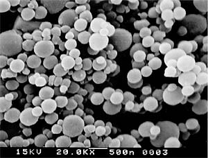
 Scanning electron microscope image of polyacrylamide beads loaded with protein — a prototype for a vaccine. Scientists hope to inject protein-loaded beads into the body, where they will be consumed by macrophages. Once inside the cell, they disintegrate and release the protein, which is then chewed up by the macrophage and the pieces displayed on the cell surface to activate T cells against the protein. Scale bar is 500 nanometers, or 200 times smaller than the width of a human hair. (Credit: Fréchet lab, UC Berkeley) |
Professor of chemistry Jean M. Fréchet, with postdoctoral fellow Niren Murthy and their colleagues, designed a polymer that falls apart in acid to form thousands of little molecules that swell and explode the cell's digestive chamber before the acids have a chance to degrade the antigens. The technique avoids a big problem of similar techniques: the cell's stomach acids often destroy the protein antigens before they can be used for display on the cell surface. Without such display, the immune system cannot detect the presence of the foreign protein.
Tests so far have been conducted only in cultured cells, but the results were impressive enough to warrant test injections in mice, which are currently underway.
Fréchet, who reported the results this week (April 21) in the early online edition of The Proceedings of the National Academy of Sciences, said the technique skirts the disadvantages of today's injectable vaccines, which employ deactivated viruses to ferry antigens into the cell interior. The antigens stimulate an immune attack against an invading organism or a cancer.
"Deactivated viruses are not always so deactivated, so it is not clear whether viruses can always be turned into a vaccine," said Fréchet. "We've developed a general delivery system that can be adapted to many different proteins. What's good about it is, it's simple."
Fréchet is head of Materials Synthesis in the Materials Science Division at Lawrence Berkeley National Laboratory and director of the Organic and Macromolecular Facility at its Molecular Foundry.
The immune system typically treats invading organisms roughly. Scavenger cells, mostly macrophages and dendritic cells, engulf and try to eat them, basically tearing them limb from limb and displaying the pieces on their surface. These scavenger cells then communicate with roving cytotoxic T cells to tell them that, should they encounter any of these pieces - called antigens - they should attack without mercy.
Vaccines are a way to prime the immune system to attack a virus or cancer without actually causing the disease. The easiest way is to disable a virus with chemicals or heat so that it can still invade cells and carry in antigens, yet not reproduce. The polio, smallpox and influenza vaccines are like this.
Other approaches include encapsulating viral or cancer antigen proteins in other materials, such as bubbles of fat called liposomes, to ferry them into scavenger cells so they will get displayed on the cell surface.
Fréchet decided to try microgel polymer beads, which are known to be snatched up by scavenger cells and degraded. Fréchet's experience in developing novel materials led him to create a bead that would, under the right conditions, fall apart into so many pieces that osmotic pressure - the tendency of water to rush in to dilute concentrations of chemicals - would draw water into the cell's digestive chamber so quickly that it would balloon and burst before the useful proteins could be degraded.
The beads he created are a new type of microgel polymer sensitive to the acidity of its environment. The chemical link holding the polyacrylamide together can be engineered to break at varying levels of acidity, and thus tuned to the acidity inside the digestive chambers, or phagosomes, of scavenger cells. (Cells engulf food in internal structures called phagosomes, which merge with acid-filled lysosomes to become highly acidic digestive chambers.)
Making the beads is a bit like making latex paint by suspension polymerization, he said. Mix the polymer chemicals with the protein antigens in a solvent - in this case, hexane - and the water loving chemicals form tiny spheres, just as oil forms globules in water. The polymer chemicals in the spheres solidify into beads with the protein embedded. His technique can produce beads of varying sizes geared to a specific use. It can produce beads - about half a micron in diameter - that are just the right size for the immune system's macrophages. Or, they can be made about a tenth of a micron in diameter to target other types of cells involved in the immune system.
To test the new method, Fréchet and his chemistry colleagues teamed up with UC Berkeley immunologist Nilabh Shastri, professor of molecular and cell biology, who developed about ten years ago at UC Berkeley a T cell assay to determine how well scavenger cells chew up and display bits of protein - so called antigen presentation. The assay indicates whether the delivery mechanisms are working and having the desired outcome, Shastri said.
The assay, which uses an egg protein, demonstrated that about 80 percent of the protein is released in about six hours under acidic conditions typical of the phagosome. In nearly neutral conditions characteristic of blood, only 10 percent is released. Scavenger cells, called antigen presenting cells, successfully displayed antigenic information on the cell surface, which would presumably allow T cells to be activated in the body as well, Shastri said.
"This is certainly an advance over previously available techniques," he said. "The beads go into the cells quite efficiently, dissolve by themselves and let go of the proteins, fragments of which end up on the cell surface in a form the T cells can recognize."
While this success is promising, animal experiments are needed to prove the method works in the body and doesn't have unsuspected side effects, Fréchet said. Nevertheless, he sees broad application for the technique in delivering proteins or genes or anti-sense RNA into cell interiors, complementing techniques that exist already.
"I'm interested in the generality of the concept," he said. "There are much flashier methods that are more complicated but probably not practical. I like the simplicity of this."
The work was supported by the U.S. Department of Energy, the National Institutes of Health, and by the Center for New Directions in Organic Synthesis, a group in UC Berkeley's College of Chemistry that is funded by Bristol-Myers Squibb and Novartis.
Fréchet's coauthors, in addition to Murthy and Shastri, are Mingcheng Xu and Stephany Schuck of the Department of Chemistry, and Jun Kunisawa of the Department of Molecular and Cell Biology.

