

UC Berkeley Press Release
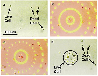 |
Selective collection of live cells from a mixture of live and dead cells: In (a), the cells are randomly positioned. In (b) and (c), a series of optically projected concentric circles round up live cells, while dead cells (stained with Trypan blue dye) leak out through the dark gaps and are not collected. The optical pattern has a yellowish color, while weak background scattered light results in a pinkish hue in the non-patterned areas. (D) shows live cells rounded up by the optoelectronic tweezer. (Images and video clips courtesy of Wu Lab, UC Berkeley) |
Engineers create optoelectronic tweezers to round up cells, microparticles
BERKELEY – Rounding up wayward cells and particles on a microscope slide can be as difficult as corralling wild horses on the range, particularly if there's a need to separate a single individual from the group.
But now, a new device developed by University of California, Berkeley, engineers, and dubbed an "optoelectronic tweezer," will enable researchers to easily manipulate large numbers of single cells and particles using optical images projected on a glass slide coated with photoconductive materials.
|
|
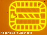
 Video image of optical "cages" created by an optoelectronic tweezer. The 20 micrometer-sized particles can be separated and sorted from those that are 45 micrometers. • Macintosh users: 1.3 Mb Quicktime video • Windows users: 6.4 Mb MPEG-4 video |
This technique, reported in the July 21 issue of the journal Nature, has an advantage over existing methods of manipulating cells, such as optical tweezers that use focused laser beams to "trap" small molecules. Such techniques require high-powered lasers, and their tight focusing requirements fundamentally limit the number of cells that can be moved at the same time.
Wu and his UC Berkeley graduate students, Pei Yu Chiou and Aaron Ohta, also improved upon other cell manipulation tools that use electrokinetic forces to create electric fields that either repel or attract particles in order to move them. Dielectrophoresis, for instance, can move larger numbers of particles. However, it lacks the resolution and flexibility of optical tweezers.
The UC Berkeley engineers found a way to get the best of both worlds by transforming optical energy to electrical energy through the use of a photoconductive surface. The idea is similar to that used in the ubiquitous office copier machine. In xerography, a document is scanned and transferred onto a photosensitive drum, which attracts dyes of carbon particles that are rolled onto a piece of paper to reproduce the image.
In this case, the researchers use a photosensitive surface made of amorphous silicon, a common material used in solar cells and flat-panel displays. Microscopic polystyrene particles suspended in a liquid were sandwiched between a piece of glass and the photoconductive material. Wherever light would hit the photosensitive material, it would behave like a conducting electrode, while areas not exposed to light would behave like a non-conducting insulator. Once a light source is removed, the photosensitive material returns to normal.
Depending upon the properties of the particles or cells being studied, they will either be attracted to or repelled by the electric field generated by the optoelectronic tweezer. Either way, the researchers can use that behavior to scoot particles where they want them to go.
Part of Wu's research was conducted while he was an electrical engineering professor at UC Los Angeles, and a co-principal investigator at NASA's Institute for Cell Mimetic Space Exploration, headquartered at UCLA's Henry Samueli School of Engineering and Applied Science.
There are many reasons why researchers would want the ability to easily manipulate cells. Biologists may want to isolate and study the fetal cells that can be found in a mother's blood sample, for instance, or sort out abnormally shaped organisms from healthy ones.
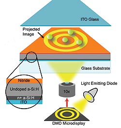 Schematic of the optoelectronic tweezer developed by UC Berkeley engineers: Liquid that contains microscopic particles is sandwiched between the top indium tin oxide (ITO) glass and the bottom photosensitive surface, made up of amorphous silicon (a-Si:H) and silicon nitride. The illumination source is a light-emitting diode operating at a wavelength of 625 nm. The optical images shown on the digital micromirror display (DMD) are focused onto the photosensitive surface and create the non-uniform electric field for manipulation of the particles. |
"Our design has a strong practical advantage in that, unlike optical tweezers, a simple light source, such as a light-emitting diode or halogen lamp, is powerful enough," said Chiou, a Ph.D. student in electrical engineering and computer sciences and lead author of the paper. "That is about 100,000 times less intense than the power required for optical tweezers."
Moreover, since the optoelectronic tweezers generate patterns through projected light, an almost limitless range of patterns are possible. Interested in boxing up individual particles in a grid-like pattern? No problem. Perhaps a star pattern would be more interesting. And there's no reason why the light needs to be static, so the researchers have even created moving conveyor belts to show how large particles can be automatically sorted from smaller ones.
"We can almost change these patterns on the fly," said Ohta, also a Ph.D. student in electrical engineering and computer sciences. "For other manipulation tools, changing these electrode patterns meant fabricating a new chip. Now, we can just project a new image to generate any type of pattern we want."
The researchers also took advantage of the difference in electrical conductivity between living and dead cells. Living cells with intact membranes in a lower conductive medium are attracted to areas of exposed light. Using a series of ever shrinking concentric circles, the researchers were able to round up living human immune cells while leaving dead ones behind.
Chiou added that while researchers can use the optoelectronic tweezer to study a few single cells, they would also have the choice of manipulating roughly 10,000 cells or particles at the same time, giving statistical weight to their studies.
The researchers are now studying ways to combine this technology with computer pattern recognition so that the sorting process could be automated. "We could design the program to separate cells by size, luminescence, texture, fluorescent tags and basically any characteristic that can be distinguished visually," said Wu.
This research is also supported by the Defense Advanced Research Project Agency, the Graduate Research and Education in Adaptive Bio-Technology training program, and the National Science Foundation.