

New findings may help improve brain-imaging tools
The minutest improvement in resolution — even a few millimeters — could help researchers better understand neural activity
![]()
| 05 March 2003
| | 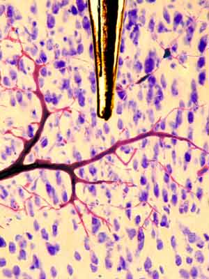 Berkeley researchers used a custom-made sensor to record both neural activity from single cells and oxygen levels from the surrounding blood vessels. They confirmed that nerve-cell activation is directly linked to an initial decrease in oxygen levels, a finding that could lead to the development of higher-resolution brain imaging methods. Image courtesy of Matthew Peterson and Michael Jacob |
New findings by Berkeley researchers could significantly improve the resolution of scans from functional magnetic resonance imaging, one of neuroscience’s most powerful research tools to date.
Functional MRI (fMRI) is a non-invasive procedure that detects increased levels of blood flow into certain areas of the brain to infer neural activity. But in a study published Feb. 14 in the journal Science, researchers from Berkeley’s Group in Vision Science show that an initial decrease in oxygen levels is an earlier and more spatially precise signal of nerve cell activity.
The findings could lead to more highly detailed fMRI scans, measured in micrometers rather than millimeters, the resolution level of most current fMRI techniques. There is hope that the new findings can provide the groundwork for research that will translate into future clinical use, such as earlier detection of brain and neurologic disorders such as Alzheimer’s or Parkinson’s diseases.
“This study has clear implications for basic neuroscience,” said Dr. Mark D’Esposito, a professor of neuroscience and psychology who is head of Berkeley’s Henry H. Wheeler, Jr. Brain Imaging Center. “A few millimeters in the brain translate into hundreds of thousands of neurons. There is no doubt that any method that can improve spatial resolution will help scientists better understand brain function.”
“All diseases begin at the cellular level,” said Ralph Freeman, professor of vision science and optometry and principal investigator of the study. “If we can eventually get a better look at what is occurring at the cellular level, then we can get a better handle on disease processes at an earlier stage in their development.”
The experiments also confirm a longstanding hypothesis that nerve-cell activation is directly connected with increased oxygen consumption at a very localized level. The researchers used a custom-made sensor that combined two microelectrodes. One electrode measured the electrical activity of a single neuron, while the other measured the concentration of oxygen in an area about 60 micrometers in diameter. The diameter of a fine human hair, in comparison, is roughly 80 micrometers.
In the experiments, the researchers monitored nerve cells in the visual cortex of four cats. Visual stimuli were displayed on a monitor in front of the cats to trigger specific neurons. They found statistically significant decreases in oxygen levels while these neurons were activated. In all, 21 nerve cells were studied.
The Berkeley study showed that when neurons are triggered, they immediately take in oxygen to fuel their activity, leading to a decrease in levels of oxygenated hemoglobin. The body reacts to this decreased oxygen level by sending a rush of oxygen-rich blood to the area.
“A common reaction among neuroscientists when they learn of this study is, ‘Why hasn’t this been done before?’” said Freeman. “Measuring metabolic and neuronal activity at the same time provides direct evidence for the link that had been theorized for so long.”
Dealing with the ‘initial dip’
Functional-MRI technology takes advantage of the fact that deoxygenated hemoglobin has slightly different magnetic properties than oxygenated hemoglobin. Machines use powerful magnets and radio waves to translate the metabolic changes into an image of brain activity.
“The initial dip in oxygen occurs in a very localized area,” said Jeffrey Thompson, a Berkeley graduate student in vision science and lead author of the study. “The subsequent increase in oxygen, which most fMRI scans measure, covers a relatively broader area. Zeroing in on the early dip could substantially improve the spatial resolution of fMRI.”
Specific challenges must be resolved before the findings are put to clinical use. For instance, because the “initial dip” of oxygen levels is relatively small, the signal is weak and will be more difficult to find.
Thompson compares the problem with finding a radio station with a weak signal. It may be possible to detect the signal by increasing the volume, but that would also lead to an increase in static and background noise that would need to be filtered out.
Nevertheless, the researchers say the study results justify further research on this initial dip in oxygen in the effort to create higher-resolution fMRI scans that can be used to better understand and detect brain disorders.
The study was also co-authored by Matthew Peterson, a Berkeley graduate student in vision science. Funding for the study was provided by grants from the National Eye Institute, part of the National Institutes of Health.