

Berkeleyan
Cell-phone video: Its not just for YouTube anymore
Researchers harness ubiquitous technology to bring medical imaging to underserved populations
![]()
| 30 April 2008
Thanks to an innovative concept developed by Berkeley engineers, cell phones could one day be used to make medical imaging accessible to billions of people around the world.
 Small file sizes mean medical-imaging data can be processed easily by a conventional cell phone. |
According to the World Health Organization, some three-quarters of the worlds population has no access to ultrasounds, X-rays, magnetic-resonance images, and other medical-imaging technology used for a wide range of applications, from detecting tumors and confirming tuberculosis infections to monitoring developing fetuses.
Medical imaging is something we take for granted in industrialized countries, says Boris Rubinsky, a Berkeley professor of bioengineering and mechanical engineering and head of the team that developed the new cell-phone application. Imaging is considered one of the most important achievements in modern medicine. Diagnosis and treatment of an estimated 20 percent of diseases would benefit from medical imaging, yet this advancement has been out of reach for millions of people in the world because the equipment is too costly to maintain. Our system would make imaging technology inexpensive and accessible for these underserved populations.
This new technique for medical imaging is described in the April 30 issue of PLoS ONE, a peer-reviewed, open-access journal published by the Public Library of Science.
Rubinsky, who holds a joint appointment as director of the Research Center for Bioengineering in the Service of Humanity and Society at Hebrew University in Jerusalem, worked on this project with Ph.D. student Yair Granot and postdoctoral researcher Antoni Ivorra. Both researchers are in Berkeleys Biophysics Graduate Group.
Rubinsky notes that simply donating imaging devices to the worlds poorest regions is not a viable long-term solution. More than half of the medical equipment in developing countries is left unused or broken because it is too complicated or expensive to operate and repair, he says. We set out to develop something that locals could sustain on their own, as well as something that is relevant to local economies and technologies.
Most medical-imaging devices consist of three essential components: the data-acquisition hardware that is connected to the patient, the image-processing software, and a monitor to display the image. When these components are combined into one unit, machine parts often become redundant, substantially increasing the cost of the device.
 Boris Rubinsky (Peg Skorpinski photo) |
Rubinsky and his team came up with the novel idea of physically separating these components so that the most complicated element the processing software used to reconstruct the raw data into a meaningful image can reside at an offsite central location, presumably in a large center where resources are available for its operation and maintenance. This central location would be used to service multiple remote sites where far simpler machines collect the raw data from the patients.
Thats where the cell phone comes in. The phone, hooked up to the data-acquisition device, would transmit the raw data to the central server, where the information would be used to create an image. The server would then relay the image back to the cell phone, for viewing on the phones screen.
This design significantly lowers the cost of medical imaging because the apparatus at the patient site is greatly simplified, and there is no need for personnel highly trained in imaging processing, says Ivorra, the postdoctoral researcher. The data-acquisition device can be made with off-the-shelf parts that somebody with basic technical training can operate. As for cell phones, you could be out in the middle of a remote village and still have cell-phone access. Theyre so prevalent because so little infrastructure is required to maintain wireless networks.
Data size ridiculously small
The principle behind medical imaging is the production of a map based upon the physical properties of different types of tissue, such as tumors, muscle, and fat. An MRI, for instance, produces a map of proton density in different tissue, while an ultrasound produces a map based upon pressure waves.
The researchers chose electrical impedance tomography (EIT) to demonstrate the feasibility of using cell phones in medical imaging. EIT is based upon the principle that diseased tissue transmits electrical currents differently than does healthy tissue. The difference in resistance to electrical currents is translated into an image.
The National Center for Research Resources at the U.S. National Institutes of Health is supporting Rubinskys research on the use of EIT to control gene therapy and cancer treatment in patients. The findings reported in the PLoS ONE paper demonstrate that these advanced medical technologies, which are dependent on EIT imaging, are not restricted to highly industrialized locations. Instead, they can be used in underserved areas of the world where there are limited resources.
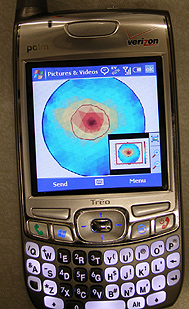
Utilizing commercially available parts, the research team built a simple data-acquisition device for the experiment. The device had 32 stainless-steel electrodes half to inject the electrical current and the other half to measure the voltage connected to a gel-filled container that simulated breast tissue with a tumor. A total of 225 voltage measurements were taken and uploaded to a cell phone, which was then dialed into a powerful central computer containing software to process the transmitted packet of raw data; an image was then reconstructed and sent back to the cell phone for viewing.
The researchers verified that the simulated tumor was clearly visible in the image, demonstrating the proof-of-principle that this system is feasible.
This could open up whole new avenues of health care for the developing world, says Rubinsky. Health professionals in rural clinics could affordably get the tools they need to properly diagnose and treat their patients.
The researchers say this system could work with any cell phone capable of sending and receiving multimedia messages such as graphics or video and audio clips. The size of the data in the study was only 6 kilobytes, which is ridiculously small, says Granot, the Ph.D. student on the research team. A one-sentence, text-only e-mail message is bigger than that.
What about dropped calls? There is no medical application that would not allow us to redial a line, says Granot. Transmitting voice signals is actually more challenging than sending this imaging data, so it shouldnt be a serious problem.
Rubinsky says the screen size of a cell phone should not be a major impediment either, in an era when people are able to watch full movies on their iPods.