Nation's
most powerful brain scanner devoted solely to research inaugurates
new era of brain research at UC Berkeley
20
Nov 2000
By
Pat McBroom, Media Relations
See
fMRI scans from a recent experiment
Berkeley
- The most powerful magnetic resonance imaging (MRI) scanner
in the country to be devoted solely to basic research on the
brain is being unveiled today (Monday, Nov. 20) at the University
of California, Berkeley.
The new
$5 million brain imaging center launches an era of extraordinary
neuroscience research at UC Berkeley. It brings together scientists
from many disciplines -physics, chemistry, biology, psychology
and computer sciences - to study the living brain with this
state-of-the-art research tool.
The MRI
scanner will be officially launched today as part of the Henry
H. Wheeler, Jr. Brain Imaging Center, where the 14-ton scanner,
manufactured by Varian, Inc., of Palo Alto, was installed
earlier this year. Inauguration of the center moves forward
UC Berkeley's Health Sciences Initiative, a commitment to
deploy the campus's rich intellectual resources across disciplines
to solve problems of human disease and unlock the mysteries
of the mind.
"We are
thrilled to officially welcome this new magnetic imaging scanner
and the Henry H. Wheeler, Jr. Brain Imaging Center to our
campus," said UC Berkeley Chancellor Robert Berdahl.
"Berkeley
is proud to lead a new era of neuroscience research and to
continue, through this important part of our Health Science
Initiative, to study the brain in an unprecedented way and
seek solutions to our most pressing health problems," he said.
Research
using the new scanner will include studies of both normal
and neurologically-impaired individuals, as scientists seek
to understand the impact of aging on memory and attention,
as well as how these functions are disturbed in people with
Alzheimer's disease, Parkinson's disease, and attention deficit
disorder.
"This is
a very special facility, one of the few in the world which
is used purely for basic research by neuroscientists, with
collaboration from physical and chemical scientists who can
push forward the frontiers of technology," said Corey Goodman,
who holds the Evan Rauch Chair of Neuroscience and leads UC
Berkeley's Helen Wills Neuroscience Institute, the parent
organization of the Brain Imaging Center.
"We want
to integrate the neurosciences from one end to the other,
from psychology and behavior to the nuts and bolts of genes
and genomes," said Goodman.
Roughly
three times more powerful than the 1.5 Tesla MRI scanners
used for clinical purposes, this research scanner can visualize
anatomical detail less than a millimeter in size. The smaller
machines can visualize detail only in the range of a few millimeters
- a major difference in terms of neural activity.
More importantly,
the machine is fast enough to support advanced work with functional
MRI (fMRI), in which neuroscientists detect and display brain
activity less than a second long. (See fMRI
scans from a recent experiment)
"Mental
events last only a few milliseconds," said Mark D'Esposito,
M.D., UC Berkeley professor of neuroscience and psychology
and director of the new Brain Imaging Center.
"Only in
the last few years have we been capable of capturing this
level of brain activity, and each year our methods improve,"
said D'Esposito. "The temporal and spatial resolution provided
by this machine will give us a unique view of neural activity
as it moves across the brain."
The non-invasive
procedure carries no risk for the individual undergoing a
scan.
Detection
of brain activity by the MRI depends on blood flow throughout
the three-pound organ. Theoretically, the flow of oxygen-rich
blood corresponds to changes in neural activity. Neurons use
the oxygen, resulting in hemoglobin changes that are detected
as radio signals by the MRI. As it turns out, deoxygenated
hemoglobin has slightly different magnetic properties than
oxygenated hemoglobin. Radio signals detected by the MRI can
then be analyzed by computer and displayed as colored areas
of the brain.
Research
already underway at UC Berkeley, using volunteers, has focused
on the brain's frontal lobes, the area just behind the forehead
that mediates so-called higher cortical functions such as
memory, attention and concentration. This area also provides
control of visual, spatial and motor activity. (See fMRI
scans from a recent experiment)
D'Esposito,
a neurologist and the first of six faculty members to be hired
by UC Berkeley's neuroscience institute, has found that, during
memory tasks, the frontal lobes function differently in young
people (ages 18-25), compared to older people (ages 60-80).
"Clearly,
short-term memory declines with age, and we see the corresponding
changes in brain activity using MRI," he said. "We can show
that the frontal lobes function differently in the two groups
during a memory task. Differences in brain activity in the
younger and older subjects was limited to one region of the
frontal lobes. In this region, older individuals used more
of the brain even though they were as accurate as the young
during performance of the task.
"This finding
suggests that, for older subjects, using more of the brain
in memory tasks has beneficial effects," said D'Esposito,
adding that there was a great deal of variation in performance
within the group.
"Why these
differences in brain activity occur between individuals and
age groups, and how this affects their performance, are clearly
key questions we have to answer," he said. "This research
has only begun."
Other kinds
of brain research with the MRI will involve subjects with
certain kinds of neurological damage who have participated
as volunteers in a wide variety of basic studies on visual
perception, motor function, memory, language and attention.
This research is being carried out by another neurologist,
Robert Knight, M.D., a professor of psychology at UC Berkeley.
Knight,
who came to UC Berkeley last year, was the first neurologist
in more than a century to be hired by a psychology department
anywhere in the nation.
Goodman,
the neuroscience institute director, said the campus's acquisition
of the new MRI scanner "exemplifies Berkeley's commitment
to health science and to integrating basic neuroscience with
medical therapy for neurological and psychiatric disorders."
Many of the researchers involved in the new brain research
are from the Department of Psychology, in the College of Letters
& Science.
###
Related
story:
Got
the picture?: A magnetic imager with three times the
sharpness of standard scanners goes online
(Berkeleyan, 27 Nov)
Links:
Health
Sciences Initiative
| |
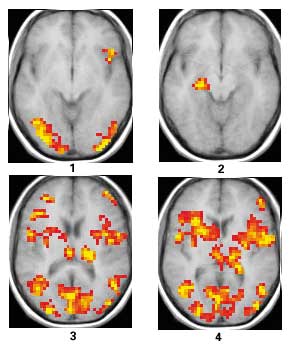
Below
are four fMRI (functional magnetic resonance imaging)
brain scans obtained during a visual memory task.
The results of the experiment were reported in November
2000 by Mark D'Esposito and Charan Ranganath of the University
of California, Berkeley, at the annual meeting of the
Society for Neuroscience. |
| |
|
| |
In
scan 1, a subject is asked to remember a face.
Areas at the rear of the brain that process visual information
are active during this task, as is an area in the frontal
lobe.
In
scan 2, the subject is asked to "think about
this face." Surprisingly, the hippocampus is activated
- the first time this has been documented. The hippocampus
was already known to be important for memory, but these
results show that this part of the brain is specifically
active during the time when we are remembering new information.
In
scans 3 and 4, the subject was asked to compare
another face to the remembered face. Some of the same
visual areas are activated as during the initial memory
task, but other areas, such as part of the frontal lobe,
are involved in making a decision about the memory.
Credit:
Mark D'Esposito and Charan Ranganath Department of Psychology
& Helen Wills Neuroscience Institute University of California,
Berkeley (2000)
|
|