|
HOME | SEARCH | ARCHIVE |
|
Got the picture?
A magnetic imager with
three times the sharpness of standard scanners goes online
![]()
By Diane Ainsworth,
Public Affairs
| |
 Peg Skorpinski photo |
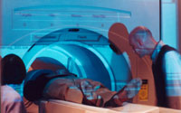
29 NOV 2000 | With its ability to capture anatomical detail a millimeter in size - smaller than the head of a pin - Berkeley's new high-resolution magnetic resonance imaging scanner will allow researchers to peer into the complex chemical and electrical circuitry of brain events that last only a few milliseconds.
The 4 Tesla imager is the only imaging instrument in the country dedicated exclusively to brain research. Using this powerful new tool to build detailed 2-D and 3-D maps of tissue types, neuroscientists, psychologists, chemists, physicists and computer scientists can probe the brain with roughly three times the magnetic strength of scanners routinely used in medical imaging.
Neuroscientists know that to watch the trillion-cell human brain in all of its stunning 3-D Technicolor - as its chemical circuitry trips local anatomic networks into thinking, reasoning, remembering and creativity - is to see the engine of human evolution spring to life. What regions of the brain come alive, for instance, when a biological switch is flipped on to coordi-nate the fingers of a concert pianist or to create a three-dimensional landscape from light that falls on a two-dimensional retina?
Probing childhood disorders
Stephen Hinshaw, a professor of psychology who specializes in child psychopathology, says the level of detail in scans of the cerebral cortex, the visual pathway, the medulla, cerebellum, corpus callosum and many other regions of the brain will unlock some of the puzzles behind such abnormalities as attention deficit disorder in children, learning problems and disturbances that put children at risk for aggression and violence.
"We will begin to localize which pathways in the brain might mediate the specific attention deficits and specific learning deficits that these kids have," he said.
Hinshaw, who also is an investigator for the National Institute of Mental Health, will be able to do this by scanning the brains of children diagnosed with these ailments while they are performing certain tasks, such as a math or memory exercise, a visual puzzle or simple physical movement. The MRI's power - 100,000 times stronger than the Earth's magnetic field - will produce laser-sharp functional maps of their brains and show researchers which pathways are stimulated in the process.
With its ability to spy 100 billion neurons in the grand syntheses of mental life itself, the magnetic scanner will illuminate specific neural networks that play key roles in this disorder. A control group of children on the normal learning curve will also undergo imaging so researchers can compare the anatomical structures and physiological processes of normal versus learning-impaired children as they perform cognitive tasks.
"We will get a very powerful look at the anatomy of these physiological processes in the brain," Hinshaw said. "But that's only part of story. By collaborating with cognitive psychologists, we'll be able to determine which tasks measure the mental processes involved in these disorders, and then we'll understand what the images mean."
Enhancing MRI sensitivity
Alexander Pines, a professor of chemistry and principal investigator of the Lawrence Berkeley Laboratory's Materials Sciences Division, plans to address problems in physics, chemistry and materials science using the 4 Tesla scanner. A pioneer in nuclear magnetic resonance spectroscopy and magnetic resonance imaging, Pines has patented technology at the Lawrence Berkeley National Laboratory, licensed recently by Nycomed Amersharm, that enhances the sensitivity of magnetic resonance imaging using xenon, an inert gas that is readily absorbed in solutions and is commonly used as an anesthetic. The technique amplifies the tiny magnetic signatures of nuclear spins and enhances the sensitivity of signal detection and associated spatial images.
Hydrogen atoms are the sole targets of magnetic resonance imaging. Although the human body is made up of untold billions of atoms, hydrogen atoms are ideal for this technique because they have a single proton and a large "magnetic moment." That is, when placed in a magnetic field, the hydrogen atom has a strong tendency to line up with the direction of the magnetic field.
Magnetic resonance imaging detects the directional spin of a hydrogen atom's nucleus, but the technique has suffered from its relative insensitivity; only one to 10 atoms out of every million spin in an "up" or "down" position and, consequently, can be detected.
Pines, together with collaborator Thomas Budinger, a medical doctor and chairman of the Bioengineering Department and their co-workers, are experimenting with xenon and some other gases that mark tissues for magnetic resonance imaging by accentuating the telltale traces of magnetism. Using a decades-old technique known as optical pumping, the investigators are able to align the nuclear spins of these magnetically marked atoms with a laser light and literally flip their spins to point upward or downward, so that they will more efficiently line up with the magnetic field and afford enhanced detection of signals. They are continuing to refine the technique, but the results are already opening the way to novel applications of nuclear magnetic resonance and magnetic resonance imaging in biomedicine.
Studying stroke, trauma
Robert Knight, a medical doctor and Berkeley professor of psychology, will use the 4 Tesla scanner to study stroke patients and victims of trauma. By imaging the flow of blood and chemical activity as nerve impulses move across and through the highly convoluted cortex, he said, researchers will be able to investigate neurological functions and connections, which allow different parts of the brain to communicate with each other.
"This technology is synonymous with getting a bigger positron smasher," he said. "It's the strength of the magnetic field that will allow us to obtain better magnetic signals and see areas of the brain with greater anatomical detail."
Related story:
Home | Search | Archive | About | Contact | More News
Copyright 2000, The Regents of the University of California.
Produced and maintained by the Office of Public Affairs at UC Berkeley.
Comments? E-mail berkeleyan@pa.urel.berkeley.edu.